When it comes to developing biologics, maintaining stability throughout the entire manufacturing process is crucial for success, which can be monitored through subvisible particle analysis. Yet, there are a number of factors that can contribute to instability and aggregation, ultimately affecting the safety and efficacy of the final product. Shear from pumping operations, suboptimal storage temperatures, freeze-thaw cycles, air-water interfaces, and exposure to various surface chemistries are just some of the culprits that can impact biologic stability.1
One factor that requires close attention is the presence of subvisible particles (SVPs). Given that SVPs have been linked to undesired immune responses, it is essential to characterize and control their presence during manufacturing. Microscopy is a method that can be used to directly count and characterize SVPs, described in both USP 7882 and Ph.Eur.2.9.19. However, manual light microscopy is low-throughput and yields poorer results than the light obscuration (LO) technique.1
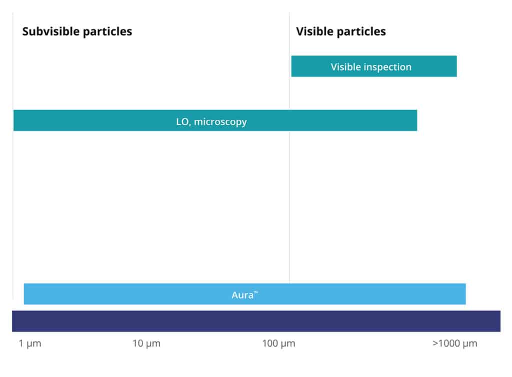
LO is currently the standard for quantifying particles ≥ 10 µm and ≥ 25 µm. However, this particle analysis method has its drawbacks, including the inability to accurately count particles in high-viscosity formulations and assess particle morphological information. In many cases, particles are transparent and not spherical, so they do not effectively obscure light and are undercounted.1
Alternatively, researchers may use flow imaging microscopy, with FlowCam® and MFI™ being the most common commercially available instruments that use this technique. This method captures images of particles flowing through a cell, enabling the calculation of size, shape, count, and other morphological features. However, issues concerning the flow cell, such as clogging and the need for cleaning pre-and post-experiment, alongside high sample volume requirements (e.g., >1 mL), remain a concern. 1
Thus, there is a pressing need for a reliable, high throughput, and low volume method for characterizing SVPs of biologics.
Flow Imaging vs. BMI: Is Backgrounded Membrane Imaging an Alternative Method for Subvisible Particle Analysis?
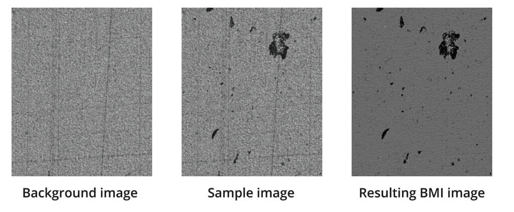
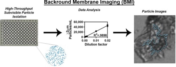
In a recent study, scientists looked at Backgrounded Membrane Imaging (BMI), which can be found on all Aura® systems, as an alternative technique. This method isolates SVPs from a liquid sample onto a filter for counting by microscope. The BMI software images the baseline before particle isolation and then subtracts that baseline from the isolated particles, leaving only photographic information from the isolate.1
Two unique protein molecules which are known to undergo aggregation as a main route of degradation were looked at by the researchers in this study. Freeze-thaw stress was applied to produce SVPs that are commonly seen throughout development. The team compared sensitivity, linearity, precision, and morphological information using LO, flow imaging, and BMI. They found that BMI performed similarly to MFI overall, while LO uniformly undercounted SVPs and had the lowest sensitivity. The sensitivity of quantification by BMI was found to be equal to or better than MFI. It is worth noting that BMI results are most aligned with FlowCam regarding both linear range and sensitivity. For example, using the trimer as an example at a dilution factor of 0.02, BMI measured 11,940 particles mL−1, and the FlowCam measured 11,690 particles mL−1. In contrast, MFI measured only 5,776 particles mL−1at the ≥2 μm size bin.1
While FlowCam had the highest magnification and most sensitive technique, it required high levels of dilution to avoid clogging the 50 μm field of view flow cell width. Moreover, the threshold parameters were optimized specifically for the enzymes tested. The low MFI counts indicate that the other two methods have a higher degree of sensitivity to small particles. While this is expected due to FlowCam’s higher magnification, it gives BMI a significant advantage, thanks to its high-throughput capabilities.
The study demonstrated that BMI offers many advantages over LO and flow microscopy techniques based on the removal of aqueous buffers from the system. Given that filter plates are single-use, there is no need to spend time rinsing or cleaning them. There is also no requirement for a pump to regulate the flow, which reduces the likelihood of calibration errors. Additionally, vacuum-mediated SVP isolation eliminates the need for degassing solutions, which can introduce air bubbles that artificially increase particulate concentrations in flow microscopy and LO techniques. Lastly, because BMI subtracts the background image, there is minimal risk of undercounting due to particle transparency.
The authors recommend employing orthogonal techniques such as BMI for particle characterization to obtain a better understanding during development. Furthermore, utilizing a high-throughput technique with low sample requirements in earlier stages of development can be very beneficial, particularly when material availability may be limited.
Use BMI for Better Particle Analysis in Biologics
BMI has been shown to be a useful tool in the measurement of protein aggregates in a high-throughput manner…
– Vargas (2020)
BMI is a superior technique that delivers comparable or better precision and sensitivity compared to current flow imaging techniques. It also presents an advantage over other SVP quantification methods, as it is high-throughput, simple, fast, and requires low sample volumes. The use of BMI in the early stages of development can save time and money while ensuring a comprehensive understanding of the product’s stability and quality.
References
- Vargas SK, Eskafi A, Carter E, Ciaccio N. A comparison of background membrane imaging versus flow technologies for subvisible particle analysis of biologics. Int J Pharm. 2020 Mar 30;578:119072. doi: 10.1016/j.ijpharm.2020.119072. Epub 2020 Jan 27. PMID: 32001293.
- https://www.uspnf.com/sites/default/files/usp_pdf/EN/USPNF/revisionGeneralChapter788.pdf


